 What happens when technology and medicine merge? Many things, one being radiology. As technology continuously evolves, it contributes to a multitude of industries in the quest to help mankind. For doctors, the key to a cure is an accurate diagnostic. But when the debilitating condition is internal, the evaluation can become tedious. The emergence of radiology centers has brought peace to medical professionals as well as patients. Through images from MRIs, CT scans, X-rays and Ultrasounds, radiology has granted greater vision to the medical field.
There are distinct differences between each of these exams but each one has the power to detect what may be deteriorating your body.
MRI scans
An MRI, acronym for Magnetic Resonance Imaging, is an exam performed with a scanner that utilizes magnetic fields and radiowaves to develop images of a human’s internal composition. Most doctors refer patients to a radiologist office when injured tendons, spinal cords or ligaments have surfaced. Brain tumors have also been detected through MRI and have been preferred for patients who may need an alternative to exam with radiation because an MRI machine does not emit any.
CT imaging
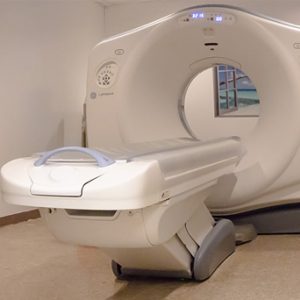
The CT scan has been helpful to physicians in detecting cancers, respiratory problems developing in the lungs and chest and has also given an in depth look into injured bones. The machine is able to develop an image quickly and displays more details compared to an MRI.
X-ray lab
Like the MRI, an X-ray evaluation is commonly used to observe broken bones and conditions like pneumonia. Instead of radiowaves, the X-ray delivers a low dose of radiation at a higher frequency intended to travel through the body in order to capture an image of your body’s internal composition.
Ultrasound imaging
For many expecting mothers, the Ultrasound is the first encounter a mother has with her unborn child. During an ultrasound, a medical professional or radiologist will use a type of scanning machine that collects sound waves to create an image of internal organs like the gall bladder, liver, kidneys, ovaries and uterus.
Early Detection Screening
You can also take charge of your health with an Early Detection Screening. Most of these exams are prescribed by a physician and then administered by a radiologist. Radiologists understand the technologies, the images developed by them and are equipped with the skills to interpret the images in order to send you and your physician accurate results.

